Dicom Images Sharing Process
Digital Imaging and Communications in Medicine (DICOM) is an imaging standard created by the American College of Radiology and the National Electrical Manufacturers Association (NEMA). The DICOM format assures that medical pictures meet high-quality criteria, allowing for an accurate diagnosis and interpretation.
Medical imaging may be viewed, edited and even reconstructed using specialist software found in all radiology departments of hospitals and clinics.
High-quality DICOM pictures have made it easier for doctors to extract as much information from a medical image as possible. Researchers are looking for ways to speed up taking a picture and applying it to patient care.
What Is DICOM?
DICOM is the standard medical imaging and information-sharing language. It stores, transmit and displays medical pictures. DICOM integration of medical imaging devices includes scanners and MRIs and how they store digital pictures.
It outlines how imaging equipment and applications may exchange pictures. Only license holders may implement the copyrighted standard.
DICOM offers a vocabulary for labeling pictures so computers and people can comprehend them. This standard applies to radiography, fluoroscopy and ultrasound imaging instruments.
https://thumbs.dreamstime.com/b/diagnostic-hospital-2142374.jpg
Why Share DICOM Images?
Medical imaging has come a long way since it was first used in the late 19th century. Today, it is used for diagnosing and treating many diseases in medical practices across the globe. However, transferring images from one country to another is not always easy.
The transfer of images in the DICOM format makes it easier for medical professionals to access them. DICOM image sharing is crucial for research, patient care and education, as it allows medical professionals to share images and reports easily.
Medical researchers can now easily share images and reports and conduct studies across different institutions hence efficient medical image management. Patient care is improved as the exchange of information is more efficient between healthcare providers.
Medical education is now available to a wider audience due to the increased access to images.
DICOM vs PACS
PACS (Picture Archive and Communication System) is a system that stores and manages medical images. It enables users to access images remotely by using the internet. A PACS system can store DICOM images. In many ways, DICOM and PACS are very similar.
Both DICOM and PACS are used to store and manage medical images. However, there are also some key differences between DICOM and PACS. The main difference between DICOM and PACS is that DICOM images can be exchanged and shared between different computers and devices.
In contrast, PACS images are only accessible from within a single facility.
The Importance of DICOM in Medical Imaging
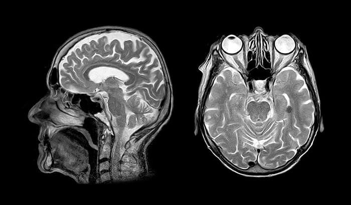
Medical imaging is an essential part of diagnosing and treating many diseases. It is used to visualize the internal organs and their blood supply, as well as the bones and soft tissue in the body. Today’s most common imaging techniques are X-rays, ultrasounds and magnetic resonance imaging (MRI).
The images taken with these techniques are vulnerable to image deterioration and distortion. Computerized image data is less vulnerable to such issues because it is already in digital format. These images are also easily transferrable and can be saved in different file formats.
DICOM is an international standard for healthcare imaging and allows for secure image share. It is also a communication protocol when moving data between PACS systems and imaging devices.
https://www.covetus.com/uploads/topics/15893746953158.jpg
DICOM Images Sharing Process
The process of sharing images begins with taking a picture. For example, a radiologist takes a picture of a patient’s knee to check for osteoarthritis. They then save the image in a DICOM server or a PACS server. The image is tagged with additional data (e.g., location and patient name).
The radiologist then shares DICOM image with medical professionals from other hospitals using FTP or a secure cloud platform. The receiving party can access the image using the DICOM viewer software.
Medical Image Sharing Use Cases
There are so many different use cases where medical images can be shared. A system that allows for easy and secure sharing of images across a network is essential for a hospital or clinic. Below are some of the most common cases where medical images are shared.
1. Consultation
A doctor may need to consult with a colleague, such as a specialist, on a patient’s condition. In this case, the specialist could view a patient’s medical images and provide their opinion on the condition.
2. Remote Locations
Remote locations, such as clinics in a remote part of the world, could share medical images with a central location. It allows specialists to provide better care to patients who may not have easy access.
3. Sharing Imaging Data
When medical professionals across the globe share images and reports, it becomes easier to diagnose and treat patients.
4. Sharing Patient Records
With DICOM images, patients no longer have to visit their doctor’s office to get their records. The doctor can print their imaging reports, put them in a folder and send them to the patient. Patients can share their records with other health care providers if they are traveling or have a new doctor.
5. Clinical Training and Education
Medical students and aspiring doctors can use the data collected from real patients to learn and practice their skills.
Medical images are essential for diagnostics and treatment. The DICOM standard ensures that all images are high quality, allowing for an accurate diagnosis. Many cloud-based image-sharing platforms have a DICOMs’ viewer built-in, making it easier for clinicians to find and use images.

